Skip to content Skip to footer

Jackson/Verbavatz Lab – The ER tether VAPA is required for proper cell motility and anchors ER-PM contact sites to focal adhesions

The Jackson/Verbavatz Lab published a new article eLife: The ER tether VAPA is required for proper cell motility and anchors ER-PM contact sites to focal adhesions   Abstract: Cell motility processes highly depend on the membrane distribution of Phosphoinositides, giving rise to cytoskeleton reshaping and membrane trafficking events. Membrane contact sites serve as platforms for direct lipid exchange…

Read More

Courtier Lab – CNCA aligns small annotated genomes

The Courtier Lab contributed to a new article published in BMC Bioinformatics : CNCA aligns small annotated genomes   Abstract: Background To explore the evolutionary history of sequences, a sequence alignment is a first and necessary step, and its quality is crucial. In the context of the study of the proximal origins of SARS-CoV-2 coronavirus, we wanted to…

Read More

JAK-STAT-dependent contact between follicle cells and the oocyte controls Drosophila anterior-posterior polarity and germline development

A new article has been published in Nature Communications: JAK-STAT-dependent contact between follicle cells and the oocyte controls Drosophila anterior-posterior polarity and germline development This study is the result of a collaboration between scientists of the Institut de biologie intégrative de la cellule (université Paris-Saclay) and Institut Jacques Monod (Konstantinides Lab, Minc Lab, Guichet Lab).   Abstract: The number of embryonic…

Read More

Pintard Lab – Microtubule-binding domains in Katanin p80 subunit are essential for severing activity in C. elegans

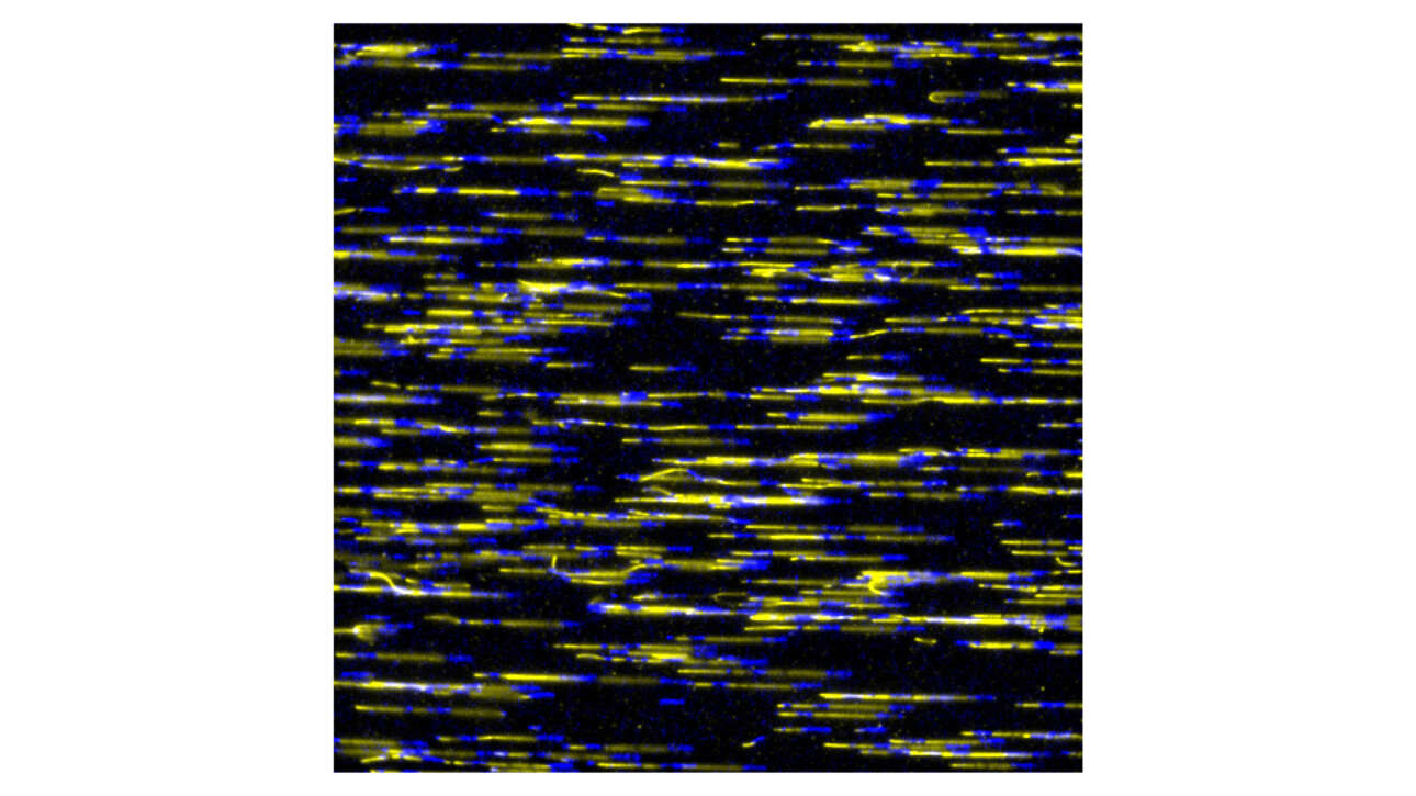
The Pintard lab recently published a new article in Journal of Cell Biology: Microtubule-binding domains in Katanin p80 subunit are essential for severing activity in C. elegans   Abstract: Microtubule-severing enzymes (MSEs), such as Katanin, Spastin, and Fidgetin play essential roles in cell division and neurogenesis. They damage the microtubule (MT) lattice, which can either destroy or amplify the…

Read More

Courtier Lab – Higher evolutionary dynamics of gene copy number for Drosophila glue genes located near short repeat sequences

The Courtier lab recently published a new article in BMC Ecology and Evolution: Higher evolutionary dynamics of gene copy number for Drosophila glue genes located near short repeat sequences Abstract: Background During evolution, genes can experience duplications, losses, inversions and gene conversions. Why certain genes are more dynamic than others is poorly understood. Here we examine…

Read More

Wassmann Lab – Qualitative rather than quantitative phosphoregulation shapes the end of meiosis I in budding yeast

The Wassmann lab published a new article in EMBO: Qualitative rather than quantitative phosphoregulation shapes the end of meiosis I in budding yeast   Abstract: Exit from mitosis is brought about by dramatic changes in the phosphoproteome landscape. A drop in Cyclin-dependent kinase (Cdk) activity, the master regulatory kinase, and activation of counteracting phosphatases such as Cdc14 in budding…

Read More

Romet-Lemonne/Jégou Lab – Cortactin stabilizes actin branches by bridging activated Arp2/3 to its nucleated actin filament

The Romet-Lemonne/Jégou Lab contributed to the publication of a new article in Nature structural & molecular biology: Cortactin stabilizes actin branches by bridging activated Arp2/3 to its nucleated actin filament   Résumé: Regulation of the assembly and turnover of branched actin filament networks nucleated by the Arp2/3 complex is essential during many cellular processes, including cell migration and…

Read More

Romet-Lemonne/Jégou Lab – Regeneration of actin filament branches from the same Arp2/3 complex

The Romet-Lemonne/Jégou Lab published a new article in Science Advances: Regeneration of actin filament branches from the same Arp2/3 complex Abstract: Branched actin filaments are found in many key cellular structures. Branches are nucleated by the Arp2/3 complex activated by nucleation-promoting factor (NPF) proteins and bound to the side of preexisting “mother” filaments. Over time, branches dissociate…

Read More